3d Mammogram Procedure
16
July
2022
What is a 3D Mammogram?
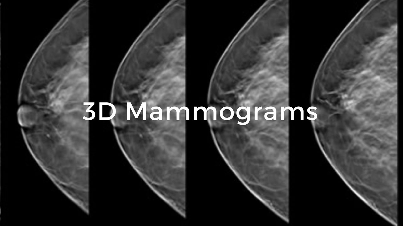
A 3D mammogram (breast tomosynthesis) is an imaging test that combines multiple breast X-rays to create a three-dimensional image of the breast.
A 3D mammogram is used to look for breast cancer in people who do not have any signs or symptoms. It could also be used to investigate the cause of breast problems, such as breast mass, pain, and nipple discharge.
When used for breast cancer screening, 3D mammogram machines create 3D pictures and standard 2D mammogram pictures. Studies show that combining 3D mammograms with standard mammograms decreases the need for additional imaging and slightly increases the number of cancers detected during screening. But more study is needed to understand whether 3D mammograms might reduce the risk of dying of breast cancer more than a standard mammogram alone.
The 3D mammogram is becoming more common, but it is not available at all medical facilities.
Why it is done
A 3D mammogram is used as a breast cancer screening test to look for breast cancer in people who do not have any signs or symptoms of the disease. It could also be used to investigate breast problems, like a suspicious lump or thickening.
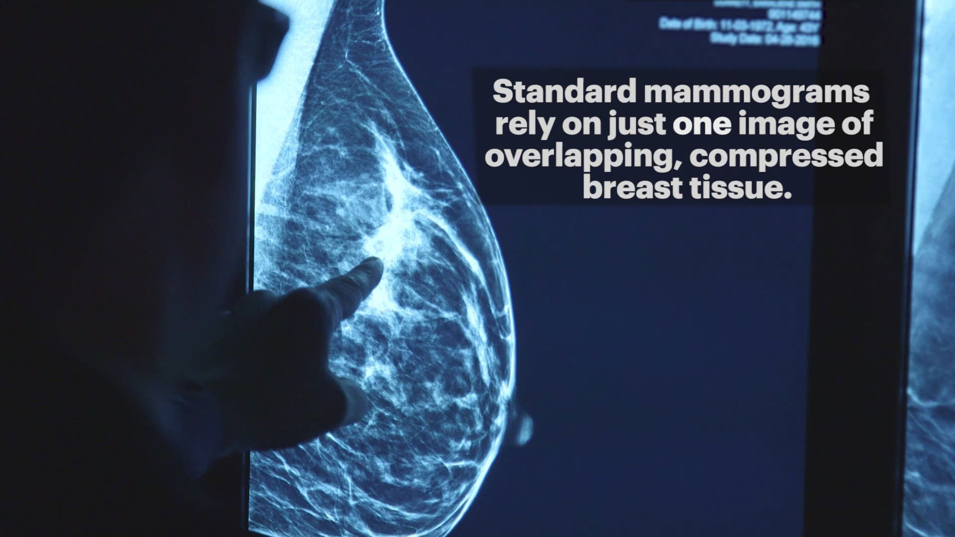
When used for breast cancer screening, the 3D mammogram machine creates 3D pictures and standard 2D mammogram pictures because both types of pictures have some advantages in seeing certain breast abnormalities.
Combining a 3D mammogram with a standard mammogram could:
- Reduce the need for follow-up imaging - When doctors detect abnormalities on standard mammogram pictures, they may recommend additional imaging. Being called back for additional imaging could be stressful. It might take extra time and lead to additional costs. Combining a 3D mammogram with a standard mammogram decreases the need for follow-up imaging.
- Detect slightly more cancers than a standard mammogram alone - Studies indicate that combining a 3D mammogram with a standard mammogram could result in about one more breast cancer for every thousand women screened when compared with a standard mammogram alone.
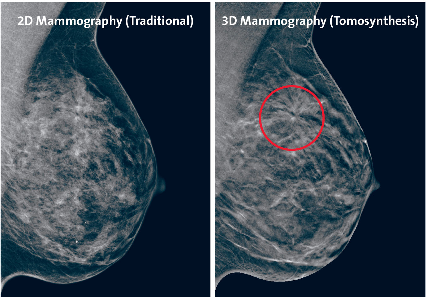
- Improve breast cancer detection in dense breast tissue - A 3D mammogram offers advantages in detecting breast cancer in people with dense breast tissue because the 3D picture allows doctors to see beyond areas of density.
Breast tissue consists of milk glands, milk ducts, supportive tissue (dense breast tissue), and fatty tissue. Dense breasts have higher amounts of dense breast tissue than fatty tissue. Both dense breast tissue and cancers appear white on a standard mammogram, which might make breast cancer more difficult to detect in dense breasts.
There is not enough evidence to conclude that 3D mammograms could reduce the risk of dying of breast cancer more than a standard mammogram alone. For this reason, most guidelines for breast cancer screening do not specify that women should choose 3D mammograms over standard mammograms alone.
3D Mammogram Risks
A 3D mammogram is a secure procedure. As with every test, it carries certain risks and limitations, like:
- Exposure to a low level of radiation - A 3D mammogram uses X-rays to create a picture of the breast, which exposes you to a low level of radiation. Because a 3D mammogram is generally combined with a standard mammogram, the level of radiation may be greater than a standard mammogram alone. Some newer 3D mammogram machines could create both 3D and 2D images at the same time, which lowers the amount of radiation.
- The test may find something that turns out to not be cancer - A 3D mammogram might identify an abnormality that, after additional tests, turns out to be benign or consistent with normal tissue. This is known as a false-positive result, and it could cause unneeded anxiety if you undergo additional imaging and testing, such as a biopsy, to further assess the suspicious area.
- The test can't detect all cancers – It is possible for a 3D mammogram to miss an area of cancer, such as if the cancer is very small or if it is in a region that is hard to see.
3D Mammogram Preparation
To prepare for your 3D mammogram:
- Choose a facility that offers 3D mammograms - Though 3D mammograms are becoming more common, they are not available everywhere. If you are interested in this test, ask your doctor whether it is available in your area.
- Check with your insurance provider - Insurance companies do not provide coverage for all 3D mammograms. Check with your insurance provider before your test so that you will know what types of costs to expect. Your insurance company might cover the standard mammogram portion of the test, while you will be responsible for the cost of the 3D mammogram portion.
- Schedule the test for a time when your breasts are least likely to be tender - If you have not gone through menopause, that is usually during the week after your menstrual period. Your breasts are most likely to be tender the week prior to and the week during your period.
- Bring your prior mammogram images - If you are going to a new facility for your 3D mammogram, gather any prior mammograms and bring them with you to your appointment so that the radiologist could compare them to your new images.
- Don't use deodorant before your mammogram - Abstain using deodorants, antiperspirants, powders, lotions, creams, or perfumes under your arms or on your breasts. Metallic particles in powders and deodorants could interfere with the imaging.
What you can expect
At the testing facility, you are given a gown and asked to remove any necklaces and clothing from the waist up. To make this simpler, wear a two-piece outfit that day.
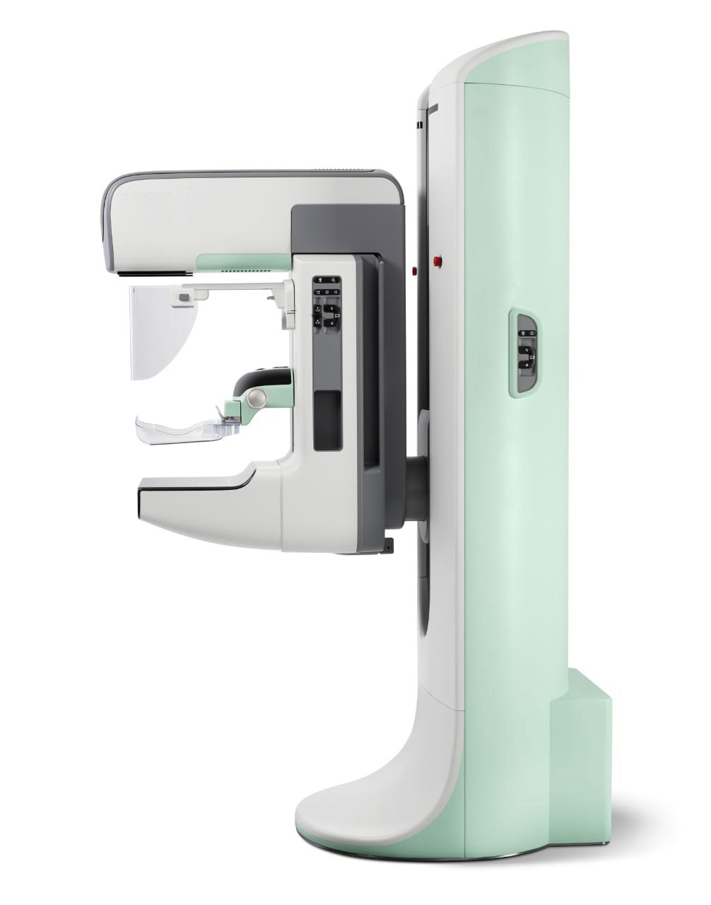
For the procedure itself, you are standing in front of an X-ray machine equipped to perform 3D mammograms. The technician puts one of your breasts on a platform and raises or lowers the platform to match your height. The technician helps you position your head, arms, and torso to enable an unobstructed view of your breast.
Your breast is slowly pressed against the platform through a clear plastic plate. Pressure is applied for a couple of seconds to spread out the breast tissue. The pressure is not harmful, but you might find it uncomfortable or even painful. If you have too much discomfort, tell your technician.
Next, the 3D mammogram machine will move above you from one side to the other as it collects pictures. You might be asked to stand still and hold your breath for a few seconds to minimize movement.
The pressure on your breast is released, and the machine is repositioned to take a picture of your breast from the side. Your breast is placed against the platform again, and the clear plastic plate is used to apply pressure. The camera takes pictures again. The process is then repeated on the second breast.
3D Mammogram Results
The pictures collected during a 3D mammogram are synthesized by a computer to form a 3D picture of your breast. The 3D mammogram pictures can be analyzed as a whole or examined in small fractions for greater detail. For breast cancer screening purposes, the machine also creates standard 2D mammogram pictures.
A doctor who specializes in interpreting imaging tests (radiologist) examines the pictures to look for abnormalities that may be breast cancer. If the radiologist sees anything unusual, he or she will use your standard mammogram and any older mammogram pictures that are available to determine whether additional testing is needed.
Additional tests for breast cancer might include an ultrasound, an MRI, or, sometimes, a biopsy to remove suspicious cells for testing in a laboratory by doctors who specialize in analyzing body tissue (pathology testing).
Hill Regional (HRH) Hospital is here to assist with all your medical needs with specialists and surgeons trained and experienced in the most advanced treatments. Our highly qualified doctors, nurses, and administrators are dedicated to caring for you with compassion in our state-of-the-art facilities.
Call us on 254-580-8500 to book an appointment with our specialist doctors.